Treatment of keratoconus
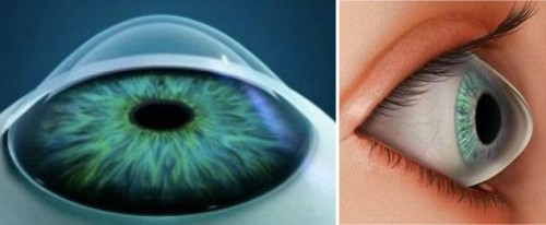
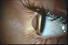
Keratoconus is a disease of the cornea, characterized by the deformation of the cornea, leading to slow but permanent thinning and improper bulging (resembling a cone protruding forward). This change in the cornea results in significant visual impairment.
Unfortunately, the exact cause of keratoconus is still unknown—it is likely a hereditary condition, and mechanical factors such as frequent eye rubbing (often associated with allergies) may also contribute to its development. It typically appears between the ages of 15 and 30 and usually affects both eyes, though to varying degrees. The progression of the disease is individual, lasting over many years, often 10, 20, or more.

Symptoms and consequences of keratoconus: worsening vision for distance, irregular astigmatism, double or shadowed vision, disorientation and reduction in distance perception, transparency disorders of the cornea.
Keratoconus cannot be completely cured, but the disease can be successfully halted and stabilized. It is often difficult to correct with glasses, although rigid contact lenses may sometimes help. In the past, corneal transplantation was the only solution. Today, transplantation can often be postponed using the Corneal Cross-linking method (CXL, CCL).
Treatment using Corneal Cross-linking (CXL, CCL)
At the Eye Centre Prague, we now offer treatment for keratoconus using the modern method of corneal strengthening—Corneal Cross-linking (CXL, CCL), which helps halt and stabilize this condition. The goal of the treatment is to strengthen the collagen bonds in the corneal tissue, stabilize it, and prevent the progression of keratoconus.
This is a minimally invasive procedure with excellent and proven results. The best outcomes can be achieved in the early stages of the disease, making timely diagnosis and prompt initiation of treatment essential.
Course of the procedure using CXL
Before the procedure, each patient must undergo a detailed examination using specialized computer diagnostic equipment and a medical examination.
|
The CXL treatment is performed on an outpatient basis, is gentle, safe, and has the ability to halt the progression of the disease, with many patients experiencing improvement in their current condition. This method can help prevent the need for corneal transplantation. |
During the CXL procedure, the eye is locally numbed with drops, making the actual procedure practically painless.
The procedure is performed while lying on a bed in the operating room. First, the epithelium of the cornea is removed. Then, riboflavin drops are applied every 2 minutes for a duration of 10 to 30 minutes. Following this, the eye is exposed to ultraviolet light for 30 minutes. By combining riboflavin (vitamin B2) with selective ultraviolet light at a wavelength of 375 nm, we create strengthening cross-links between individual molecules in the corneal tissue (similar to the process your dentist uses to harden a composite dental filling). This makes the corneal tissue stronger and more resilient. The entire procedure takes approximately 45-60 minutes. After the procedure, a contact lens is applied to the eye, which will be removed during the follow-up examination.
Treatment of keratoconus using CXL is currently not covered by health insurance (only patients insured by VZP have the procedure covered by public health insurance).


