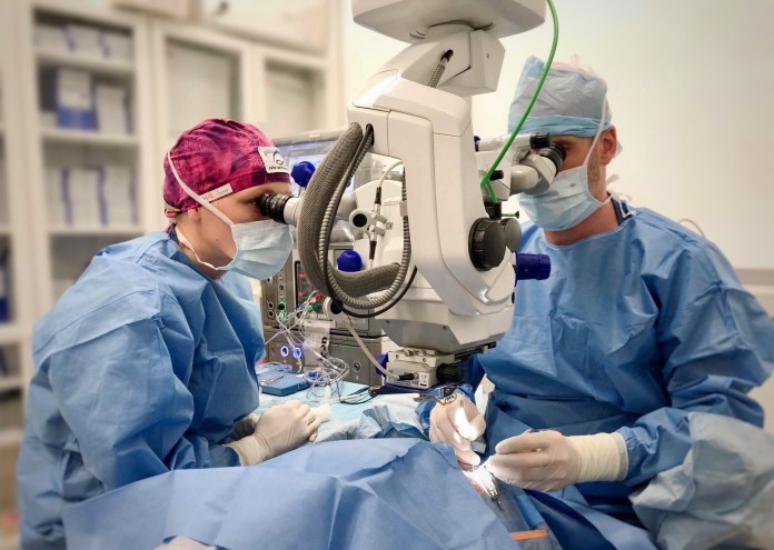
Retina and vitreous surgery – Pars plana vitrectomy (PPV)
Surgery using the Pars plana vitrectomy (PPV) technique is named after the location through which the surgeon enters the eye to perform the necessary procedures on the retina and vitreous. It is a very important and often vision-saving operation. The procedure is performed on an outpatient basis under local anesthesia, and the patient can go home afterward.
Indications for the procedure
The surgery is always preceded by a specialized examination, including OCT angiography. At the Eye Centre Prague, the recommendation to perform surgery using the PPV technique is given for the following retinal and vitreous diseases:
- Diseases at the vitreous-retina interface: macular hole, epiretinal membrane, vitreomacular and vitreoretinal traction syndrome
- Retinal diseases: macular edema, retinal detachment, diabetic retinopathy
- Vitreous diseases: vitreous floaters, vitreous inflammation and injuries, vitreous hemorrhage
The "Pars plana vitrectomy" procedure is fully covered by all health insurance providers at the Eye Centre Prague.
Procedure overview
The surgery is outpatient and takes approximately 30 minutes. It is painless—before the procedure, the eye is numbed with drops, and an injection of anesthetic is administered behind the eye.
Pars plana vitrectomy is a complex surgical procedure performed in an operating room under a microscope. The surgeon enters the eye with thin instruments through three micro-incisions and performs the necessary interventions inside the eye. During the procedure, the vitreous is removed, and the affected areas of the retina are treated with laser or freezing (cryocoagulation). At the end of the procedure, the vitreous is replaced with infusion fluid or an internal tamponade—an expanding gas or silicone oil. The internal tamponade is used when pressure needs to be applied to the retina to ensure it heals in the correct position.
Thanks to a special self-sealing technique, the incisions usually do not require stitches. The eye is covered with a sterile patch until the next day. After the surgery, the patient goes home with a companion.

Follow-up and postoperative care
The day after the procedure, a postoperative check-up will take place, during which a specialist will examine the surgical site to assess the postoperative findings.
Positioning after surgery
In some cases, after retina and vitreous surgery, the doctor may recommend specific positioning to ensure the proper placement of the gas or oil filling in the eye.
There are three head positioning options: 1. face-down position, 2. face-down with the head resting on the right cheek, 3. face-down with the head resting on the left cheek.

The specific head position is determined by the doctor for each patient. Positioning is done with approximately 5-10 minute breaks. The patient should avoid lying on their back.


